
Table of contents
Lacrimal Infections
This section covers dacryoadenitis (inflammation of the lacrimal gland), canaliculitis (inflammation of the canaliculi), and dacryocystitis (inflammation of the lacrimal sac).
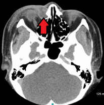
Dacryoadenitis
Dacryoadenitis is inflammation of the lacrimal gland.
Pathology
- Often idiopathic.
- Secondary causes include viral infections like mumps, Epstein-Barr virus, and herpes zoster.
Diagnostics
Presentation
- Acutely painful upper lateral lid
- Swollen lacrimal gland
- Hypoglobus (medially)
- Enophthalmos
- S-shaped eyelid deformity
Investigations
Only if recurrent or suspicious
- Orbital MRI/CT
- Biopsy
Bilateral dacryoadenitis should raise suspicion of sarcoidosis
Management
- Oral NSAIDs or steroids.
Resolution can take months
Canaliculitis
Inflammation of the canaliculi.
Pathology
- Often caused by infection, most commonly by Actinomyces
Presentation
- Unilateral epiphora and discharge
Management
- Topical antibiotics and canaliculotomy
Canalicular repair following canalicular trauma is with a Mini Monoka tube for 3 months
Dacryocystitis
Inflammation of the lacrimal sac. It requires urgent management to prevent the spread of cellulitis.
Pathology
- Caused by nasolacrimal duct obstruction, usually by Staphylococcus species
Presentation
- Presents acutely with epiphora and a tender lacrimal sac.
Management
Acute
- Warm compress and oral antibiotics.
- Incision and drainage may be required for abscess formation.
Chronic/recurrent
- Dacryocystorhinostomy (DCR) (open connection between the lacrimal sac and middle nasal meatus).
- This can be performed via external or endoscopic approach.