
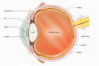
The conjunctiva is a thin transparent membrane that lines the surface of the sclera and the underside of the eyelid. It can be thought of as a modified layer of skin.
Anatomy

Conjunctival Segments
The conjunctiva is anatomically divided into 3 sections:
- Palpebral conjunctiva - lines the posterior surface of the eyelids.
- Bulbar conjunctiva - lines the anterior surface of the sclera.
- Forniceal conjunctiva - a folded layer between the palpebral and bulbar conjunctiva. It allows movement of the eyelids.
The conjunctiva does not cover the cornea. It fuses with the sclera at the limbus.
Innervation
- Main - CNV1 (ophthalmic division of the trigeminal nerve).
- Inferior conjunctiva - infraorbital nerve.
- Limbus - long ciliary nerve (branch of the nasociliary nerve).
Lymphatics
- Medial conjunctiva - submandibular nodes.
- Lateral conjunctiva - preauricular nodes.
Signs of Conjunctival Disease
Sign |
Description |
|---|---|
Hyperaemia (conjunctival injection) |
Enlargement of conjunctival vessels |
Chemosis (conjunctival oedema) |
Transparent swelling of the conjunctiva |
Conjunctival membranes |
Exudative adherences of the conjunctiva |
Cicatrization |
Scarring of the conjunctiva |
Follicles |
Discrete lesions which appear like transparent grains of rice. No vessels inside the lesion |
Papillae |
Lesions confined to the palpebral conjunctiva with a vascular center. |