
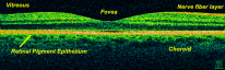
The retina is the ocular structure that is ultimately responsible for vision. The vitreous is a gel-like matrix that fills the bulk of the eye. The vitreous adheres closely to the retina and disorders of the vitreous affect the retina, and vice versa.
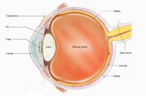
Retina
The retina can be thought of as a direct extension of the brain. It is the out-sprouting of the optic nerve that reacts to light stimuli. There are 3 main types of retinal neurons: photoreceptors, bipolar cells and ganglion cells.
Dimensions
- Thinner temporally
- Surface area of 1250mm²
- Internally bound by vitreous
- Externally bound by Bruch's membrane of the choroid
- Anteriorly continues with the outer pigmented choroidal epithelium (at the ora serrata)
Blood supply
- Inner 2/3 of retina → Central retinal artery
- Outer 1/3 of retina → Choroidal vessels
- Vessels of the retina lack internal elastic lamina
- Venous drainage is by central retinal vein into the cavernous sinus
- There are 2 blood plexuses within the neurosensory retina
- Inner plexus lies on the ganglion cell layer
- Outer plexus lies on the inner nuclear layer
The cilioretinal artery forms an anastomosis between the central retinal artery and choroidal vessels. It arises from the posterior ciliary vessels
Blood retinal barrier
- Inner blood retinal barrier : tight endothelial junctions of retinal vessels
- Outer blood retinal barrier: zonulae occludentes (Latin for tight junctions) of RPE
Retinal surface
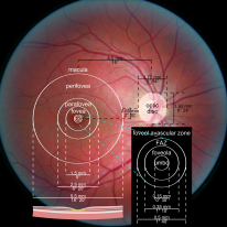
Macula
- 1.5mm
- It has greater >1 ganglion cell layers compared to the peripheral retina which only has 1
Foveal avascular zone
- 0.5mm
- Avascular and depends on supply from the choriocapillaris via diffusion
Foveola
- Fovea centralis (foveola) : 0.35mm
- No rods but the maximum concentration of cones
The blind spot is at the optic disc (contains no photoreceptors) and is 1.8mm²
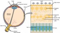
Retinal structure
2 principal components: the inner neurosensory retina and the outer retinal pigment epithelium (RPE).
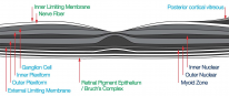
Layers of the Neurosensory retina
- Internal limiting membrane
- Nerve fiber (contains ganglion cell axons)
- Ganglion cell layer (contains cell bodies of ganglion cells)
- Inner plexiform
- Inner nuclear (contains cell bodies of glial cells and bipolar cells)
- Outer plexiform
- Outer nuclear (contains cell bodies of photoreceptors)
- External limiting membrane
- Photoreceptors
RPE
- The retinal pigment epithelium (RPE) lies outer to the photoreceptors
- A single layer of cuboidal epithelium which maintains the photoreceptors
- Bruch's membrane of the choroid lies outer to the retinal pigment epithelium
The neurosensory retina has 9 layers. The RPE is 1 layer. In total, the retina is considered to have 10 layers.
Photoreceptors
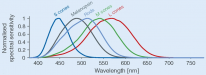
There are 2 main types of photoreceptors: rods and cones. Cones also come in 3 further subtypes
Rods |
Cones |
|---|---|
Rod number rises towards fovea and suddenly becomes 0 |
Cones have their highest concentration in fovea but 99% of the total number is outside the fovea. |
The main visual pigment in rods is rhodopsin, and it is responsible for night, dim and peripheral vision.` |
The visual pigment in cones comes in three types and are responsible for trichromatic colour and central vision
Blue cones are not found in the fovea |
Ratio of rods: cones is 20:1
Retinal neuromodulator cells
- These cells modulate action potentials
- Horizontal cells : between photoreceptor and bipolar cells
-
Amacrine cells : between bipolar and ganglion cells
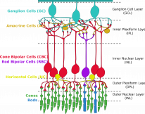
Schematic diagram of the retinal cells. By Jörg Encke, CC BY-SA 4.0.
Ratio of photoreceptors to RPE cells is 45:1
Vitreous
The vitreous is a viscoelastic gel that fills the vitreous chamber - a 4ml cavity that forms the body of the eye. It is adherent to the retina and the ora serrata.
- This gel is composed mainly of water, but also contains hyaluronic acid and type II collagen
- It is connected to the internal limiting membrane of the retina by the collagen fibrils
The vitreous gel progressively becomes runnier with age. This is called syneresis and leads to small pools of fluid within the gel. Syneresis can lead to posterior vitreous detachment from the retina.