
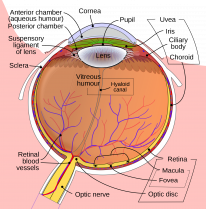
The uvea is composed of 3 parts: the choroid, iris and ciliary body. These parts are considered together as the uveal tract. The sclera is the white part at the front of the eye, wrapping all the way to the back and terminating at the optic nerve.
Choroid
The choroid is a highly vascular, pigmented structure extending from the ora Serrata to the optic disc. It provides vascular supply and absorbs reflecting light. The choroid gets thinner as you move anteriorly.
Layers of the Choroid
From external to internal:
- Haller's layer (large vessels).
- Sattler's layer (medium vessels).
- Choriocapillaris (capillaries).
- Bruch's membrane (basement membrane).
The suprachoroidal space is a potential space that sits on top of Haller's layer. It separates the sclera from the choroid.
Iris
The iris is the anterior part of the uveal tract. It extends from the anterior chamber angle to the pupil. Its muscles are responsible for changing the size of the pupil. The iris is the coloured part of the eye when you're looking at someone directly.
Dimensions
- 12mm diameter.
- 37mm circumference.
- 2mm thickness.
- The anterior chamber volume is 200µL compared to the posterior chamber at 60µL.
Iris Zones
- When looking at someone's iris head-on, the visible anterior border is described in 3 zones.
- The inner pupillary zone contains the sphincter pupillae muscle.
- The outer ciliary zone contains the dilator pupillae muscle.
- The collarette is the middle zone used for anatomical division.
Muscles
- The sphincter pupillae is responsible for pupil constriction and is innervated by postganglionic parasympathetic short ciliary fibres of CN3.
- The dilator pupillae is responsible for pupil dilations and is innervated by sympathetic branches of the ciliary nerves.
Blood Supply
- Major circle comprised of 2 long posterior and 7 anterior ciliary arteries.
- Minor circle is formed at the level of the collarette.
- Iris vessels are not fenestrated, they do not leak during FFA.
Layers of the Iris
- Anterior border.
- Stroma.
- Dilator pupillae.
- Posterior pigment epithelium.
- The anterior border is composed of modified stromal cells with crypts.
- Koganei are clumps of pigmented macrophages found in the iris stroma.
- The sphincter pupillae muscle lies within the stroma.
- The posterior pigmented epithelium is cuboidal.
Ciliary Body
A muscular secretory structure involved in accommodation, aqueous production, and the blood-aqueous barrier. Its functions are described in more detail in glaucoma and neuro-ophthalmology chapters. This section focuses more on anatomy.
Divisions
- Anterior functional part → pars plicata.
- Posterior non-functional part → pars plana.
There are around 70 major ciliary processes in the pars plicata which are involved in aqueous secretion.
Snow banking is deposition on the pars plana of the ciliary body, a characteristic clinical sign of uveitis.
Layers of the Ciliary Body
- Epithelium, stroma and muscles.
- The epithelium is bilaminar and cuboidal.
- The outer layer is pigmented.
- The inner layer is non-pigmented and produces aqueous.
- The bilaminar epithelium is arranged apex-to-apex.
- There are 3 muscles: longitudinal (outermost), oblique, circular (innermost).
Nerve Supply
- Parasympathetic: Edinger Westphal nucleus of CN III → ciliary ganglion → short ciliary nerves → contract ciliary body and laxes zonular fibres.
- Sympathetic: Hypothalamus → spinal cord → superior cervical ganglion → ICA plexus or by joining V1 → long ciliary nerves → relax the ciliary body and tenses zonular fibres.
Sclera
The sclera stretches from the iris at the front of the eye, to the optic nerve at the back. It is a tough outer coat of connective tissue. The episclera is another layer of connective tissue which sits on top of the sclera. It is highly vascular and supplies the sclera with nutrients. These 2 structures will be discussed in this section.
Sclera
- Thickest at the optic nerve.
- Thinnest posterior to rectus muscle insertions.
- Forms 5/6ths of the outer layer of the globe. The cornea forms the remainder.
- Contains mainly type 1 and 3 collagen.
- The sclera is separated from the uvea by the suprachoroidal space.
- Innervated by long and short ciliary nerves.
Episclera
- Heavily vascularised.
- The episclera is in between the sclera and the conjunctiva.
Scleritis results in inflammation of the deep vascular plexus of the sclera. This is too deep to be affected by topical phenylephrine (vasoconstrictor). Contrastingly, episcleritis leads to the inflammation of the superficial vascular plexus and is affected by topical phenylephrine. This means that the reddened vessels in episcleritis blanch with the administration of topical phenylephrine, compared to in scleritis where they don't. This is an important clinical differentiator.