
Ocular trauma can be serious and sight-threatening. It requires urgent assessment and management. The different types of ocular trauma are summarised on this page.
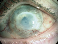
Intraocular Foreign Bodies (IOFB)
All patients with penetrating eye trauma should be investigated for IOFB. Some are highly toxic and can lead to severe disease.
Pathology
Copper
- Pure copper is the most toxic heavy metal to ocular tissues in common use.
- Alloys of copper can lead to Sunflower cataracts and Kayser-Fleischer rings (the signs of Wilson's disease).
Other bodies
- Iron deposits in the ocular epithelial tissue and lead to cell death, resulting in glaucoma, cataract and retinal detachment.
- Glass, plastic, stone, silver, gold, platinum and lead are all inert.
- Aluminium and zinc have mild toxic effects.
Investigations
- CT/X-ray should be taken in all cases.
- MRI is contraindicated as some bodies can be magnetised.
- Electroretinogram (ERG) testing will show a reduced b-wave in some cases of chronically retained IOFB.
Management
If there is a penetrating injury then primary repair of the globe is the priority. Foreign body removal technique depends on the location.
Location |
Procedure |
|---|---|
Corneal |
26G needle |
Anterior chamber |
Fine forceps |
Angle of the anterior chamber |
Scleral trapdoor approach |
Ciliary body |
Electromagnetic removal |
Posterior segment |
Intraocular magnet or vitrectomy forceps |
Burns
Thermal/chemical injuries are severe and can be blinding. Prompt treatment is essential.
Pathology
- Alkali injury is significantly worse than acid.
- Alkalis cause liquefactive necrosis.
- Acids cause coagulative necrosis.
Corneal healing is achieved by the migration of limbal stem cells. If the limbus is damaged then healing is hindered.
Management
Treatment steps
- Irrigation with copious amounts of water.
- Antibiotics + cycloplegics + lubricants + analgesia.
- Topical steroids.
- Ascorbic acid - aids collagen formation and mops up free radicals. Should not be used in acid burns!
- Doxycycline - proteinase inhibitor which aids healing.
Prolonged corneal healing problems
- Amniotic membrane transplant.
- Limbal stem cell transplant.
Topical sodium citrate inhibits collagenases and aids healing. However, administration is painful and so it is not in common use.
Orbital Floor Fractures
Orbital floor fracture is a classic traumatic ocular injury and is very likely to be tested in the exam.
Pathology
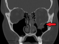
- Classically caused by ball/fist injury to the eye.
- Soft tissue of the globe is pushed into the maxillary sinus.
- The inferior rectus can be entrapped, causing vertical diplopia and restriction of eye elevation.
Surgical emphysema can also occur in orbital floor fractures but is more pronounced in medial wall fractures.
Diagnostics
Presentation
- Periorbital bruising.
- Vertical diplopia and extraocular muscle entrapment.
- Enophthalmos.
- Infraorbital anaesthesia.
Investigations
- CT is diagnostic.
- Hess charts are used in monitoring and follow-up.
In extraocular muscle entrapment, the antagonistic movement is restricted. I.e if the superior rectus is trapped, downgaze will be restricted.
Management
- Surgery does not have to be immediate, if at all necessary.
- Indications for immediate surgery include: oculocardiac reflex and severe facial destruction.
- If there is only minimal diplopia and enophthalmos, observation can suffice.
Advise patients to avoid blowing their nose.
Retrobulbar Haemorrhage
The bony orbit is a rigid chamber and intraorbital bleeding/swelling can lead to a sharp rise in pressure. This is an ophthalmic emergency and can result in blindness.
Pathology
- Trauma causes oedema/bleeding in the orbit and leads to a rise in intraorbital pressure.
- Rising intraorbital pressure restricts blood flow and causes optic nerve damage.
- The process can be thought of as orbital compartment syndrome.
Diagnostics
Presentation
- Painful loss of vision.
- Rigid proptosis.
- Restricted ocular movements.
- Relative Afferent Pupillary Defect (RAPD).
- Elevated intraocular pressure.
Investigation
- Radioimaging can aid diagnostics but treatment should not be delayed.
Management
Immediate
IV mannitol + IV acetazolamide + IV methylprednisolone.
No improvement to medications or severe
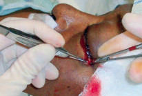
Lateral canthotomy and cantholysis.
Still no improvement
Orbital decompression.
Cantholysis is the disinsertion of the lateral canthal tendon. This procedure allows the release of intraorbital pressure.
Hyphema
Hyphema is blood in the anterior chamber and is most commonly caused by blunt trauma.
Pathology
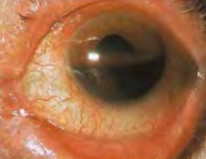
- Hyphema is a sign of ocular trauma and warrants further investigation.
- Microhyphema is when the erythrocytes are only visible on a slit lamp.
Diagnostics
- Check drug history for anticoagulants.
- Check medical history for coagulative disorders and sickle cell.
Management
Treatment is only required in high-risk cases such as those in children, rebleeds and risky medical histories.
Treatment
- Admit for steroids and bedrest.
- Monitor IOP for red cell glaucoma.
- No improvement → AC paracentesis.
Trabeculectomy - if all else fails.