
The majority of the tear film is secreted by the lacrimal gland. The drainage of these tears is facilitated by the nasolacrimal system.
Disorders of lacrimation lead to either a watery eye or a dry eye.
Anatomy and Physiology
Tear Flow
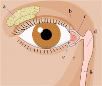
- Secretion by the lacrimal gland onto the ocular surface.
- Channeled medially by the orbicularis pump mechanism.
- Drainage into the nasolacrimal system via the upper and lower puncta.
- Flow through the upper and lower canaliculi into the common canaliculus.
- Common canaliculus → Nasolacrimal sac → Nasolacrimal duct → inferior nasal meatus.
Disorders of tearing are caused by an imbalance between secretion and excretion of tears.
Tear Film
There are 3 layers of tear film:
Lipid layer
- The thinnest and most superficial layer.
- Produced by the meibomian gland (Sebaceous).
- Prevents evaporation of the tear film.
Aqueous layer
- Produced by the lacrimal gland.
- Thickest layer.
- Has an immune function.
Mucin layer
- Deepest layer.
- Produced by conjunctival goblet cells.
- Spreads the film evenly and keeps it stable on the ocular surface.
Reflex tearing is a common cause of hypersecretion. It is caused by irritation of the ocular surface. A good quality tear film reduces reflex tearing.
Glands of Lacrimation
Lacrimal Gland (main)
- Sensory innervation → lacrimal nerve (branch of CNV1).
- Lacrimation (secretomotor) → Parasympathetic (CN7).
Accessory Lacrimal Glands
-
Krause and Wolfring glands are accessory lacrimal glands that maintain a basal aqueous layer.
- Krause glands are found at the conjunctival fornices and are more abundant in the upper fornix.
- Wolfring glands are less numerous but bigger. They are found at the tarsal plate.
Eyelash Associated Glands
- The glands of Moll and Zeis both service eyelash follicles.
- Moll glands are apocrine (modified sweat glands).
- Zeis (and Meibomian glands) are holocrine (sebaceous).
Special Tests
There are a variety of special tests to investigate the lacrimal system. These tests are detailed in the table below.
Description |
Interpretation |
|
|---|---|---|
Schirmer’s test |
Filter paper placed under the lower lid. Amount of moisture measured after 5 mins |
|
Tear film break-up time |
Eye is stained with fluorescein dye and the time taken for the first dry spot to appear on the cornea is measured |
|
Jones 1 test |
Dye squirted onto the conjunctiva and cotton bud placed in the inferior meatus. |
|
Jones 2 test |
Dye syringed into the nasolacrimal system and cotton bud placed in the inferior meatus. |
|
Dacryocystography |
Radiological evaluation using injected fluorescent contrast to evaluate the nasolacrimal system morphology |
|
Dacryoscintigraphy |
Radiological evaluation of nasolacrimal system drainage using radiopharmaceutical eyedrops |
|
The Jones tests have been largely superseded by more useful radiological imaging methods such as dacryocystography in clinical practice.